Blog
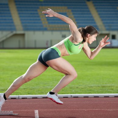
Keep it Cool and beat the Heat- Focus on Tokyo 2020.
With the Tokyo 2020 (2021) Olympics in full swing, I thought we would share some insight into cooling strategies for the athletes. This research was published in June 2020 in the British Journal of sports medicine. I have linked the article at the bottom. The conditions for athletes, like Guernseys own Cameron Chalmers, competing in Tokyo […]

Running into problems?
During the various lockdowns that have been occurring due to the pandemic, a lot of people have taken to running in order to stay fit. Unfortunately, since then, there has also been an increase in running injuries coming into our clinic. It seems perfectly reasonable to a lot of people that even if it’s been […]
Tips for working from home
Elbows, knees and hips should be at approximately 90 degrees. Monitor should be at a height that your line of sight falls in the top third of the screen. Monitor should be approximately an arm span away from you. Keep your arms relaxed at your sides, do not elevate your shoulders. Feet should be flat […]

5 key factors to consider when looking at nutrition and arthritis.
(1) Maintain a healthy body weight Excessive body weight puts extra stress on the joints, exacerbating arthritis. Adipose (fat) tissue releases hormones and chemicals into the body, some of which promote inflammation, which in turn can contribute to joint damage. (2) Increase foods rich in omega-3 fats Omega-3 fats can reduce inflammation and be protective […]
Are you waking up with neck pain?
Waking up with neck pain and stiffness, this is definitely not the way you want to start your day. You go to bed intending to get a solid 7 hours sleep and hope to wake up feeling refreshed. Here’s a few factors that may be contributing to your neck pain: Your Pillow – Your head […]

Osgood Schlatter’s Disease
What is it? OS is really a syndrome rather than a disease and is one of the most common causes of knee pain in growing children. It can be extremely painful and debilitating and generally affects active children between the ages of 9 and 16. It presents as a painful and often swollen lump at […]

Ankle Sprains
Acute Phase: Day 1 to 4 You probably think you know a lot about ankle sprains. It’s a very common topic but for a good reason. Ankle sprains are the most common injuries and account for 25% of all sports injuries. Over a series of blogs, I will take you through the different stages of […]

Shockwave Success!
Here is a great testimonial from one of our patients who had fantastic results with Shockwave Therapy. We’ve had our machine for over a year now and we are really excited about the great results we’ve had in a variety of conditions including shin splints, plantar fasciitis, shoulder tendinopathy / calcification and tennis elbow. ‘In […]

Avoiding Injury Over Christmas
BE CAREFUL OUT THERE! Unfortunately, at this time of the year, we see a rise in injuries caused by anything from the wet and icy conditions on the roads to lifting Christmas trees from storage etc. So in order to avoid your festive season being marked by pain, we thought we would offer a few […]

Winter Feet
Our feet form the foundation for our entire body so it is important to keep them healthy all year round. During the colder winter months feet are generally covered up and out of sight as we try to keep warm with thick socks, slippers and boots. However out of sight should not mean out of […]
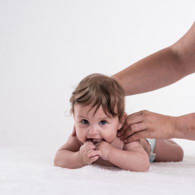
Tummy Time
What is Tummy Time? Placing your baby on their front to play when they are AWAKE and SUPERVISED! Babies should still be placed on their backs to sleep! Importance of Tummy time Provides new sensory experiences, increases physical challenges, and provides much needed relief from constant pressure to the back of the head. It also helps strengthen neck, […]

Preparing for and Recovering from Major Surgery – My ‘Hip-Hop’ Experience
Having recently undergone a full hip replacement, I am now recuperating at home with a bit of spare time on my hands. I thought it may be of interest to share some of my experience with you. I have included some do’s and don’ts that I have found useful, but obviously, each person will have […]
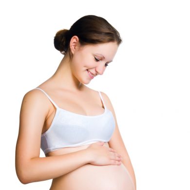
Taking Care of Yourself After Childbirth
After you hold your bundle of joy in your hands. It is easy to get carried away taking care of them that you forget to take care of yourself. Remember, only if you are in good health are you able to give your best to your child and family.
Joint Care During Winter
Simple lifestyle changes can help manage existing conditions and prevent the development of joint pain

How to help your child use more words and sentences
We see lots of children who are understanding what is being said to them, but are not yet using many words. Here are a few ideas to encourage children to begin using spoken words and phrases. How can I help my child to use words? Get face to face – when playing with your child […]
Supporting Attention and Listening Skills in Young Children
What is attention and listening? Attention and listening is being able to listen and focus on specific tasks or sounds. It can be a tricky skill for young children to learn, but underpins so much of their learning. Good attention and listening skills help with: Social skills Understanding language Following instructions Learning to communicate Speech […]
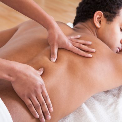
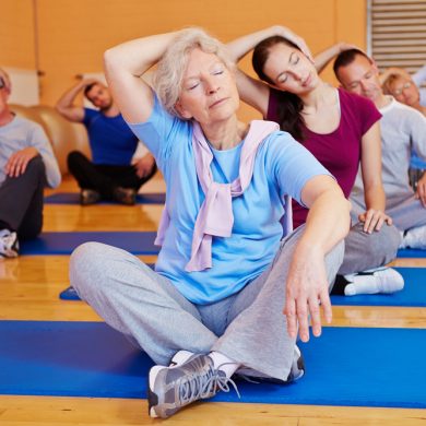
Let Massage help you boost your Immune System this winter
A study found that participants receiving regular massage saw measurable improvements to their immune systems

Core Strength and Injury Prevention
What do you ask yourself when you’re told you have a ‘weak core’ and this is why you are in pain.
What does having a ‘weak’ core mean? How does poor core stability predispose to injury?

Core training for injury prevention part 2
How to improve your core stability. Looking at common mistakes made wen core training either to rehabilitate an existing injury or prevent future injuries for people prone to suffer from back pain.

Game, Set and Tennis Elbow
As the tennis season comes into full swing and you are inspired by Murray’s or Serena’s recent wins to step onto the court. I thought it maybe the right time to talk to you about one of the most common injuries faced by tennis players and has therefore gained the name ‘Tennis Elbow’. Lateral Epicondylitis […]

Have You Heard Sitting is The New Smoking!
Okay so I will show you a picture – take a deep breath and decide is this you? Well, is it? If it is – as my dearly departed grandfather used to say “You sit like a sack of potatoes.” At this point, you may raise your eyebrows and think ‘I didn’t know that […]

Runner’s Knee
So now the sun is out, you’ve finally found the motivation to get out on the road and start running. Maybe you’ve even gone a step further and signed up for that upcoming half marathon or marathon. After putting in all this hard work the last thing you need is to be stopped in your […]

Understanding Hypermobility
Hypermobility is described as relative increased mobility to what is expected. It can occur at one joint, one movement or generalized to the whole body. This can occur as a compensation due to lack of movement at another joint, helping achieve the gross required movement. When joints are too mobile the instinctive response is that the […]
5 WAYS TO IMPROVE YOUR POSTURE AT WORK
With a large number of us working with computers in desk-based roles and spending a large amount of our free time on laptops, tablets and smartphones, It comes as no surprise that many of us start to develop what is termed upper cross syndrome (anterior head posture). This can lead to painful conditions such as […]
Shin Splints
Shin splints also known as ‘Medial tibial stress syndrome’ is a common condition which affects athletes, runners, tennis players etc. It can present as pain along the inner edge or front of the shin bone and can be a dull ache that intensifies during and after exercise. Causes include: Poor core strength, inflexibility, muscular imbalance leading […]
World Psoriasis Day 29th October 2015
World Psoriasis Day is a day specifically dedicated to people who have psoriasis and/or psoriatic arthritis. Approx 125 million people are living with psoriasis. What is Psoriasis? Psoriasis is an autoimmune disease which causes raised, red, scaly patches to appear on the skin. Psoriasis can appear in any location but particularly affects the elbows, knees […]
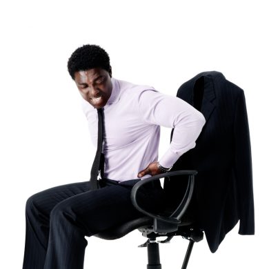
Back awareness week 2015
Back Pain and Children It was Back awareness week last week (5th to the 11th October), and the focus this year was back pain experienced by children. Prevention begins at school New shocking statistics: One quarter of UK secondary school pupils suffer from regular or daily back pain School bag burden was associated with a […]
Tips to prevent Tennis injuries
Maybe a few of you have decided to pick up your tennis racquet after the excitement of Wimbledon. I just thought I would give you a few tips on how to prevent tennis injuries. Be prepared for your sport. You could have an excellent serve and great backhand, but if you are not wearing the […]
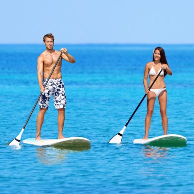
Stand Up Paddleboarding
So some of you may have experienced an unfamiliar site down the beach this summer already. YES, people standing up on boards using a paddle? Well, this is the new craze of Stand Up Paddleboarding or ‘SUP’ing for short. This relatively new watersport has been taking the world by storm over the last few years […]
Cycling: Injuries and Prevention
Following on from our World Cup Football theme, I thought I would write a small piece about cycling to keep in tune with the current Tour de France: Cycling is the third most popular recreational activity in the UK. An estimated 3.1 million people ride a bicycle each month. Grabbing your bicycle and heading out for a ride regularly […]
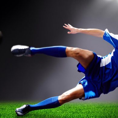
Football: Injuries and prevention
FIFA World Cup – As this is one of the most exciting times for football enthusiasts I thought a blog about football would be most suited. Football injuries are common due to the stop-start nature/speed of the game, the multi-directional stresses applied to the body and sometimes the unpredictability of what other players do. […]
Summer Footwear aka ‘ The Flip Flop! ‘
Summers arrived and the weathers here to stay, HOPEFULLY!!! Anyway, this can only mean one thing, more time outside and lots more hours clocked up wearing our beloved FLIP FLOPS!!! (Slaps, Thongs, whatever you want to call them) So apart from the obvious lack of protection making us more vulnerable to dropped objects, stubbed toes […]
Are you using the right pillow?
We lie down for about 1/3 of our lives. The secret to a good nights sleep is in neutral spinal alignment. If your pillow is too low or too high, you will not be correctly positioned. By ensuring that you are lying on a bed and pillow that is helping your alignment you are investing in your […]

Achilles Problems
The achilles tendon connects the calf muscle to the heel bone and this allows us to go up on to our tip toes and push off when walking or running. Common achilles problems include: Achilles tendonitis – this is inflammation of the tendon. Achilles tendinosis – this is when there are tiny micro tears […]
General Knee Pain
Although there are many more serious conditions that can occur at the knee joint, if you are suffering from achy knees during or post exercise and don’t recall any specific injury, you may be suffering from ‘Runners Knee’. As the name suggests, this is a common ailment amongst runners but can effect anyone who does […]
Tennis Elbow
Tennis elbow is a common condition that causes pain around the outside of the elbow and is clinically known as Lateral Epicondylitis. It is often caused through overuse of the muscles of the forearm and can cause pain during everyday activities such as: – Lifting/Bending of the arm – Gripping small objects such as a […]
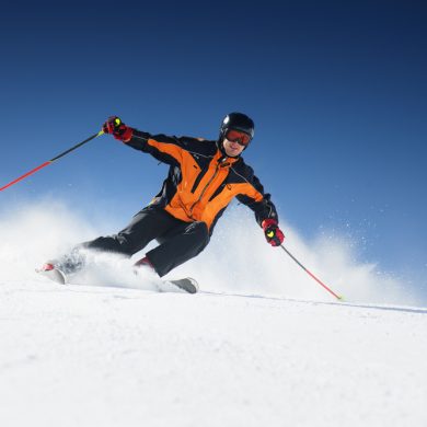
Skiing/Snowboarding injuries and prevention
Thousands of skiers and snowboards enjoy the snow-sports every year; of these very few prepare for the physical demand these sports place on the body. We take a look at common injuries, their causes and prevention. Free Downloadable skiing/snowboarding preparation programme.
Good Lifting technique: Tips to help prevent back injuries
Before you lift an object think through the task. Consider the risks factors and decide where you are going to place the object and how you will get it there. Make sure you clear a path. Get as close to the load/object as possible. Your feet should be placed approximately a shoulder width apart, one […]
Are you sitting in the optimum position?
Recently I have been questioning patients about the set up of their chair in the office. When sat in your chair are you: leaning forwards, sitting upright or leaning slightly backwards? Many studies have been carried out to analyse the optimum sitting position. It is well known that sitting upright for hours on end […]
A guide to choosing the right mattress and pillow
Do you have sleepless nights due to neck, low back and hip pain? Have you ever considered that your pillow or mattress may be the cause of the problem. Sleep is essential to our everyday functioning, so it is important that we get a good night’s rest. In order to do this you should make […]
The wonders of reflexology for fertility, pregnancy and beyond…
The following appeared on the website www.netmums.com. Reflexologist, Jacqui Booth discusses the wonders of reflexology for fertility, pregnancy and beyond… How is reflexology helpful to women trying for a baby? Reflexology is a natural, non-invasive healing art that uses pressure points on the feet and hands to balance out other parts of the body. It aims […]
Should I use Ice or Heat on an Injury?
Both heat and cold are beneficial in reducing pain, but they work in different ways. Through my experience some people are still not sure when they should be using cold or when they should be applying heat. I hope you find the following useful. Ice packs should be used on acute injuries, for example an […]
Golf: Injuries and prevention
Low back pain, wrist and elbow problems are the most common injuries seen in golfers. Despite being classified as a low-impact sport, approximately 40% of amateur and 60% of professional golfers suffer from injury each season. These injuries majority of the time are due to overuse (such as repetitive bending and twisting), poor conditioning and […]
Arthritis and Osteopathy
What is arthritis? Arthritis is a term which is used to describe inflammation of a joint. There are two main categories of arthritis: inflammatory arthritis and degenerative/mechanical arthritis (osteoarthritis). Osteoarthritis (OA) is the most common form of arthritis in the UK. Each year in the UK 9 million people seek help from their doctor for […]
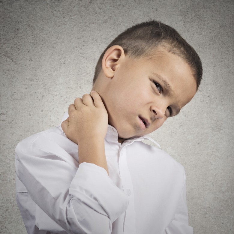
Is your child’s school bag and shoes causing them pain and possibly problems in the future.
The Back Health Charity BackCare have highlighted that approximately 60% of children are carrying over weight school bags. Some primary school children are routinely carrying bags which weigh over 15% of their body weight (NHS, 2012). Not only are the bags too heavy, most children are wearing them in the most detrimental way, on one […]
Horse riding: the influence between rider and horse
As an Osteopath and horse rider myself, I have treated many horse riders and have seen how injuries or imbalances in the rider can have an effect on the horse. If a rider if suffering with back or hip pain etc, he or she will undoubtedly be compensating for this in their riding position […]
Exercise during Pregnancy and Post-Birth
Healthcare providers are encouraging women to exercise moderately during their pregnancy, unless they have any complications or have been advised not to. Exercise before and during pregnancy has been associated with reduced preeclampsia risk, prevention of gestational diabetes and has also been linked to a decrease in caesarean section rates and infants with higher Apgar […]
Shin Splints
Shin splints Shin splints is a general term used to describe pain at the front of the lower leg. However true shin splints cause pain on the inside of the shin and this can arise from a number of causes. The pain from shin splints tends to be dull at first, but if ignored can […]
Heel pain – About Plantar Fasciitis, Prevention and Treatment
Plantar Fasciitis: how to prevent and treat it What is plantar fasciitis? Plantar fasciitis is inflammation of you plantar fascia, which is found on the sole of your foot, attaching from your heel bone to your toes. The plantar fascia is a strong band of tissue and its main job is to support the arch […]
Do You Know The Correct Height For Your Bike Saddle?
A lot more of you have been getting out on your bikes, now the weather has cheered up a bit. But do you know if your bike is set up correctly? Adjusting the height of the saddle has been shown to be important for both performance and injury prevention. The recommended height of the saddle […]
We now give you the best Kinesiology taping system in the world – Rocktape
Hi all! Andre, Dimi, Gemma and Raj spent the sunniest day of the year so far, in a dingy room with the guys from Rocktape. We learnt the secrets behind how to use this high-tech Kinesiology tape and are now officially adding Rocktape to our list of specialities. To find out a little more about […]